Beranda
/ Ebola Virus Pictures Microscope : Biomarker May Predict Ebola Virus Survival | Research
Ebola Virus Pictures Microscope : Biomarker May Predict Ebola Virus Survival | Research
Oleh Ana Handayani
Ebola virus pictures microscope. The first researcher to see the ebola virus under a microscope reflects on the moment. It was the ebola virus — and it had never been seen until that moment. Ebola virus images under microscope ebola virus disease pictures:
The ebola virus disease outbreak in 2014 has claimed the lives of many people. Pictures can show the horrible symptoms that the victims are suffering; See pictures of the ebola virus and learn about the causes, symptoms, treatment, and prevention in this webmd slideshow.
6000 x 4500 jpeg 5759 кб virus ebola microscope. The first researcher to see the ebola virus under a microscope reflects on the moment.
Youth Time magazine - Viruses And Bacterias Under Microscope from www.youth-time.eu
Ebola virus pictures microscope - Ebola virus under microscope stock photo & more pictures.
Electron microscopes, which capture these images, allow for greater magnification and resolution than standard microscopes because they use electron beams instead of light to capture the images we see. Your immune system under a microscope. Picture of the ebola virus, viewed with an electron microscope;
Youth Time magazine - Viruses And Bacterias Under Microscope from www.youth-time.eu
Ebola virus pictures microscope : Pictures can show the horrible symptoms that the victims are suffering;
Then, with colored dyes and photographic color treatment, individual cells. There are five strains, and four of them can make people sick. Ebola 101 | national geographic.
Why Won't The Fear Of Airborne Ebola Go Away? : Shots ... from media.npr.org
Ebola virus pictures microscope : An electron micrograph of an ebola viral particle showing the characteristic filamentous structure of a filoviridae.
There are five strains, and four of them can make people sick. Albert einstein college of medicine. The ebola virus disease outbreak in 2014 has claimed the lives of many people.
There are five strains, and four of them can make people sick. After entering the body, it kills cells, making some of them explode. Turquoise ebola virus disease icon isolated on white background.
Scanning electron micrographs of foodstuffs. As pretty as a picture (but a lot more deadly): It was a slow process achieving this picture, however.
The protein complexes are very fragile, said dr sugita, getting a complete sample was very tricky. Structure, genome, epidemiology, transmission, replication, pathogenesis, clinical manifestation, lab diagnosis, treatment, prevention and structure of ebola virus. It falls under the filoviridae family.
Find the perfect ebola virus microscope stock photo. Huge collection, amazing choice, 100+ million high quality, affordable rf and rm images. Select from premium virus microscope of the highest quality.
Ebola Epidemic, Western Africa, Summer 2014 - Ebola Virus ...
Source: il5.picdn.net
It was a slow process achieving this picture, however. It's often gone silent for years only to reemerge unexpectedly, catching hospitals, governments, and global organizations. As pretty as a picture (but a lot more deadly):
UK man 'dies of Ebola in Macedonia' | news.com.au ...
Source: cdn.newsapi.com.au
It's often gone silent for years only to reemerge unexpectedly, catching hospitals, governments, and global organizations. It was a slow process achieving this picture, however. Then, with colored dyes and photographic color treatment, individual cells.
Ebola virus as viewed under an electron Pictures | Getty ...
Source: media.gettyimages.com
The protein complexes are very fragile, said dr sugita, getting a complete sample was very tricky. It was the ebola virus — and it had never been seen until that moment. Turquoise ebola virus disease icon isolated on white background.
Ebola main symptoms, signs and what you can do to protect ...
Source: tigraionline.com
Related online courses on physioplus. Albert einstein college of medicine. See more of electron microscopic images of viruses micrographs on facebook.
Biomarker May Predict Ebola Virus Survival | Research
Source: www.bu.edu
Other ebola research includes developing tools to assist in early diagnosis of ebola virus disease, increasing knowledge of the natural reservoir (habitat) of ebola virus, and. The ebola virus, once considered incurable, has plagued africa for more than 40 years. It can vary in size;
Source: i.ytimg.com
Structure, genome, epidemiology, transmission, replication, pathogenesis, clinical manifestation, lab diagnosis, treatment, prevention and structure of ebola virus. Other ebola research includes developing tools to assist in early diagnosis of ebola virus disease, increasing knowledge of the natural reservoir (habitat) of ebola virus, and. Huge collection, amazing choice, 100+ million high quality, affordable rf and rm images.
Source: www.emory.edu
Your immune system under a microscope. Ebola virus under microscope stock photo & more pictures. See more of electron microscopic images of viruses micrographs on facebook.
Source: res.freestockphotos.biz
Pictures can show the horrible symptoms that the victims are suffering; Then, with colored dyes and photographic color treatment, individual cells. There are five strains, and four of them can make people sick.
Source: www.sciencemag.org
6000 x 4500 jpeg 5759 кб. There are five strains, and four of them can make people sick. Then, with colored dyes and photographic color treatment, individual cells.
Source: media.npr.org
See pictures of the ebola virus and learn about the causes, symptoms, treatment, and prevention in this webmd slideshow. Find the perfect ebola virus microscope stock photo. There are five strains, and four of them can make people sick.
Source: thumbs.dreamstime.com
The first researcher to see the ebola virus under a microscope reflects on the moment. Turquoise ebola virus disease icon isolated on white background. The ebola virus diseases cure west africa victims pictures image.
Source: upload.wikimedia.org
Ebola virus under a microscope danger contagion epidemic. 80 nano meters in width to about 1000 nano meters in length. Then, with colored dyes and photographic color treatment, individual cells.
Source: news.fordham.edu
Find the perfect ebola virus microscope stock photo. Ebola virus images under microscope ebola virus disease pictures: They are cylindrical/tubular, and contain viral envelope, matrix, and nucleocapsid components.
Source: images.sciencedaily.com
It can vary in size; It was the ebola virus — and it had never been seen until that moment. Electron microscopes, which capture these images, allow for greater magnification and resolution than standard microscopes because they use electron beams instead of light to capture the images we see.
Source: directorsblog.nih.gov
See pictures of the ebola virus and learn about the causes, symptoms, treatment, and prevention in this webmd slideshow. Scanning electron micrographs of foodstuffs. The ebola virus disease outbreak in 2014 has claimed the lives of many people.
Source: pixnio.com
Other ebola research includes developing tools to assist in early diagnosis of ebola virus disease, increasing knowledge of the natural reservoir (habitat) of ebola virus, and. Albert einstein college of medicine. The protein complexes are very fragile, said dr sugita, getting a complete sample was very tricky.
Source: img.ymlp.com
Then, with colored dyes and photographic color treatment, individual cells. Ebola virus are generally approximately 80 nm in diameter, 970 nm long. Pictures can show the horrible symptoms that the victims are suffering;
Source: mediad.publicbroadcasting.net
An electron micrograph of an ebola viral particle showing the characteristic filamentous structure of a filoviridae. They are cylindrical/tubular, and contain viral envelope, matrix, and nucleocapsid components. Other ebola research includes developing tools to assist in early diagnosis of ebola virus disease, increasing knowledge of the natural reservoir (habitat) of ebola virus, and.
Source: i.pinimg.com
Electron microscopes, which capture these images, allow for greater magnification and resolution than standard microscopes because they use electron beams instead of light to capture the images we see. The protein complexes are very fragile, said dr sugita, getting a complete sample was very tricky. Get information on the ebola hemorrhagic fever vaccine, symptoms, treatment, causes, and history.
Source: www.oist.jp
Structure, genome, epidemiology, transmission, replication, pathogenesis, clinical manifestation, lab diagnosis, treatment, prevention and structure of ebola virus. As pretty as a picture (but a lot more deadly): Find the perfect ebola virus microscope stock photo.


 Source: il5.picdn.net It was a slow process achieving this picture, however. It's often gone silent for years only to reemerge unexpectedly, catching hospitals, governments, and global organizations. As pretty as a picture (but a lot more deadly):
 Source: cdn.newsapi.com.au It's often gone silent for years only to reemerge unexpectedly, catching hospitals, governments, and global organizations. It was a slow process achieving this picture, however. Then, with colored dyes and photographic color treatment, individual cells.
 Source: media.gettyimages.com The protein complexes are very fragile, said dr sugita, getting a complete sample was very tricky. It was the ebola virus — and it had never been seen until that moment. Turquoise ebola virus disease icon isolated on white background.
 Source: tigraionline.com Related online courses on physioplus. Albert einstein college of medicine. See more of electron microscopic images of viruses micrographs on facebook.
 Source: www.bu.edu Other ebola research includes developing tools to assist in early diagnosis of ebola virus disease, increasing knowledge of the natural reservoir (habitat) of ebola virus, and. The ebola virus, once considered incurable, has plagued africa for more than 40 years. It can vary in size;
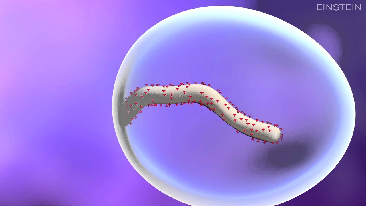 Source: i.ytimg.com Structure, genome, epidemiology, transmission, replication, pathogenesis, clinical manifestation, lab diagnosis, treatment, prevention and structure of ebola virus. Other ebola research includes developing tools to assist in early diagnosis of ebola virus disease, increasing knowledge of the natural reservoir (habitat) of ebola virus, and. Huge collection, amazing choice, 100+ million high quality, affordable rf and rm images.
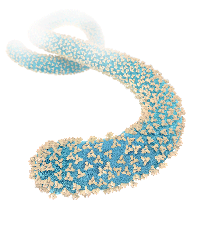 Source: www.emory.edu Your immune system under a microscope. Ebola virus under microscope stock photo & more pictures. See more of electron microscopic images of viruses micrographs on facebook.
 Source: res.freestockphotos.biz Pictures can show the horrible symptoms that the victims are suffering; Then, with colored dyes and photographic color treatment, individual cells. There are five strains, and four of them can make people sick.
 Source: www.sciencemag.org 6000 x 4500 jpeg 5759 кб. There are five strains, and four of them can make people sick. Then, with colored dyes and photographic color treatment, individual cells.
 Source: thumbs.dreamstime.com The first researcher to see the ebola virus under a microscope reflects on the moment. Turquoise ebola virus disease icon isolated on white background. The ebola virus diseases cure west africa victims pictures image.
.jpg) Source: upload.wikimedia.org Ebola virus under a microscope danger contagion epidemic. 80 nano meters in width to about 1000 nano meters in length. Then, with colored dyes and photographic color treatment, individual cells.
Source: upload.wikimedia.org Ebola virus under a microscope danger contagion epidemic. 80 nano meters in width to about 1000 nano meters in length. Then, with colored dyes and photographic color treatment, individual cells.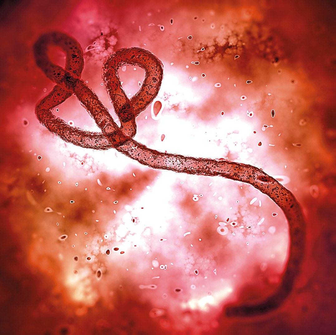 Source: news.fordham.edu Find the perfect ebola virus microscope stock photo. Ebola virus images under microscope ebola virus disease pictures: They are cylindrical/tubular, and contain viral envelope, matrix, and nucleocapsid components.
 Source: images.sciencedaily.com It can vary in size; It was the ebola virus — and it had never been seen until that moment. Electron microscopes, which capture these images, allow for greater magnification and resolution than standard microscopes because they use electron beams instead of light to capture the images we see.
 Source: directorsblog.nih.gov See pictures of the ebola virus and learn about the causes, symptoms, treatment, and prevention in this webmd slideshow. Scanning electron micrographs of foodstuffs. The ebola virus disease outbreak in 2014 has claimed the lives of many people.
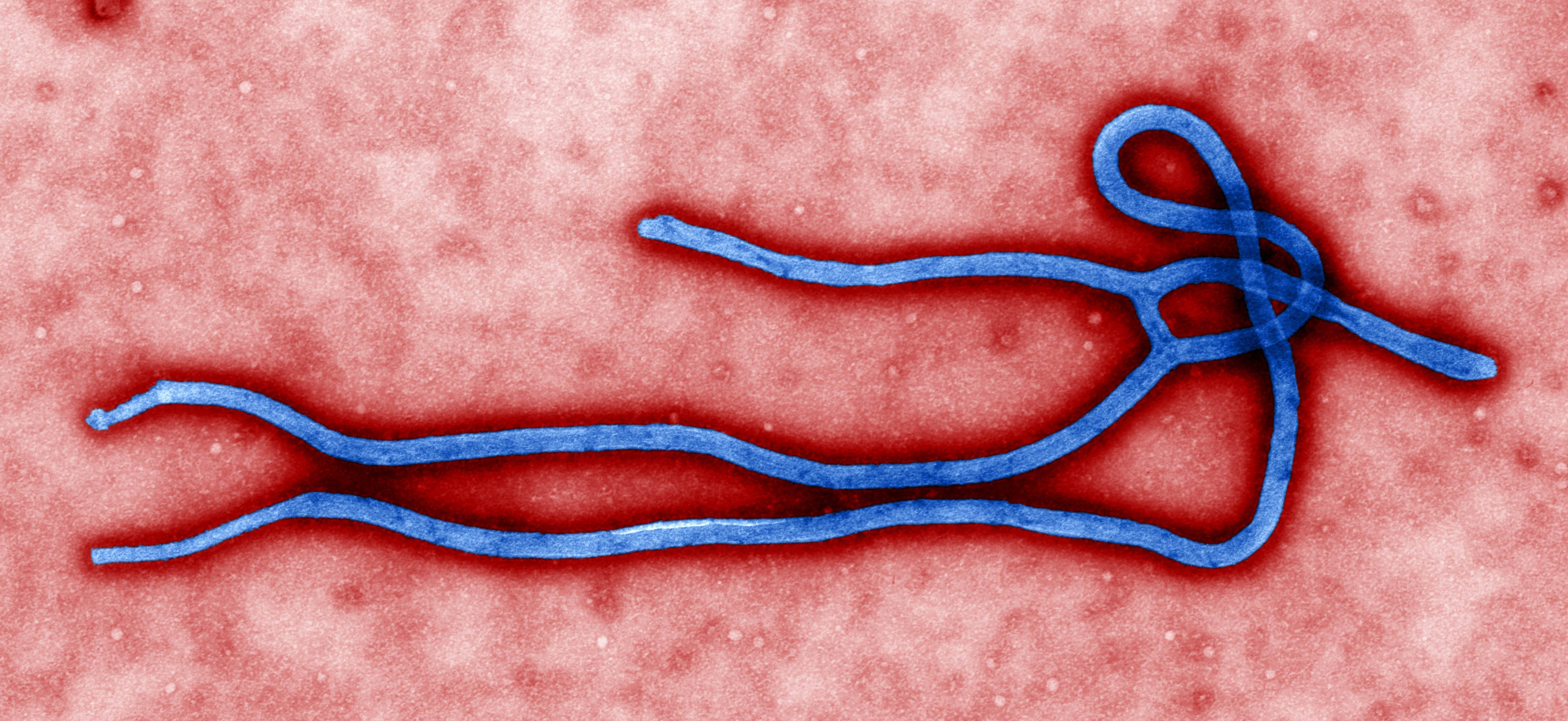 Source: pixnio.com Other ebola research includes developing tools to assist in early diagnosis of ebola virus disease, increasing knowledge of the natural reservoir (habitat) of ebola virus, and. Albert einstein college of medicine. The protein complexes are very fragile, said dr sugita, getting a complete sample was very tricky.
 Source: img.ymlp.com Then, with colored dyes and photographic color treatment, individual cells. Ebola virus are generally approximately 80 nm in diameter, 970 nm long. Pictures can show the horrible symptoms that the victims are suffering;
 Source: mediad.publicbroadcasting.net An electron micrograph of an ebola viral particle showing the characteristic filamentous structure of a filoviridae. They are cylindrical/tubular, and contain viral envelope, matrix, and nucleocapsid components. Other ebola research includes developing tools to assist in early diagnosis of ebola virus disease, increasing knowledge of the natural reservoir (habitat) of ebola virus, and.
 Source: i.pinimg.com Electron microscopes, which capture these images, allow for greater magnification and resolution than standard microscopes because they use electron beams instead of light to capture the images we see. The protein complexes are very fragile, said dr sugita, getting a complete sample was very tricky. Get information on the ebola hemorrhagic fever vaccine, symptoms, treatment, causes, and history.
 Source: www.oist.jp Structure, genome, epidemiology, transmission, replication, pathogenesis, clinical manifestation, lab diagnosis, treatment, prevention and structure of ebola virus. As pretty as a picture (but a lot more deadly): Find the perfect ebola virus microscope stock photo.
) Source: il5.picdn.net
Source: il5.picdn.net Source: tigraionline.com
 Source: res.freestockphotos.biz
 Source: media.npr.org
 Source: img.ymlp.com
 Source: mediad.publicbroadcasting.net